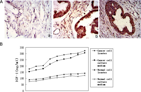
Figure 1.

Level of IGF-1R and IGF-1 in EOC. (A) IGF-1R expression in surgical specimens of epithelial ovarian cancer (EOC). Representative pictures of surgical samples with different IGF-1R expression (original magnification ×100). (a) IGF-1R protein expression was low in benign epithelial serous ovarian tumor tissues. (b,c) EOC tissues clearly expressed IGF-1R protein. Magnification was in small square. (B) Quantitated IGF-1 level in tumor cell lysates and cell culture medium by ELISA. IGF-1 level in tumor cell lysates and cell culture medium is much higher than in normal epithelial ovarian cell lysates and cell culture medium(n=10)(P<0.05). [2 independent sample tests].
