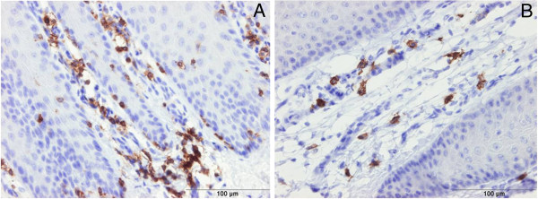
Figure 3.

Immunohistochemical staining of digital dermatitis infected skin for detection of CD3 (+ve T-lymphocytes ) (A, M0) and CD20 (+ve B-lymphocytes) (B, M4). T and B lymphocytes were observed mainly in the dermis at the junction with the epidermis.
