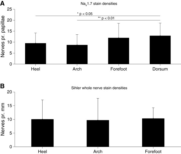
Figure 3.
Quantification of the identified nerves from Nav 1.7 immunoreactivity and Sihler staining. A– Number of intrapapillary nerves per papillae showing NaV1.7 immunoreactivity. There were significant differences between the sites (1-way ANOVA, p < 0.01, F(3,170 = 5.174); the heel (post-hoc, p < 0.05) and arch (post-hoc, p < 0.01) had significantly lower nerve fiber densities than the dorsum. There were no statistically significant differences between the sites in the sole of the foot. B– No significant differences in the densities of intradermal nerve fibers were found between the sites stained with Sihler’s method (1-way ANOVA, p=0.994, F(2,8) = 0.006).

