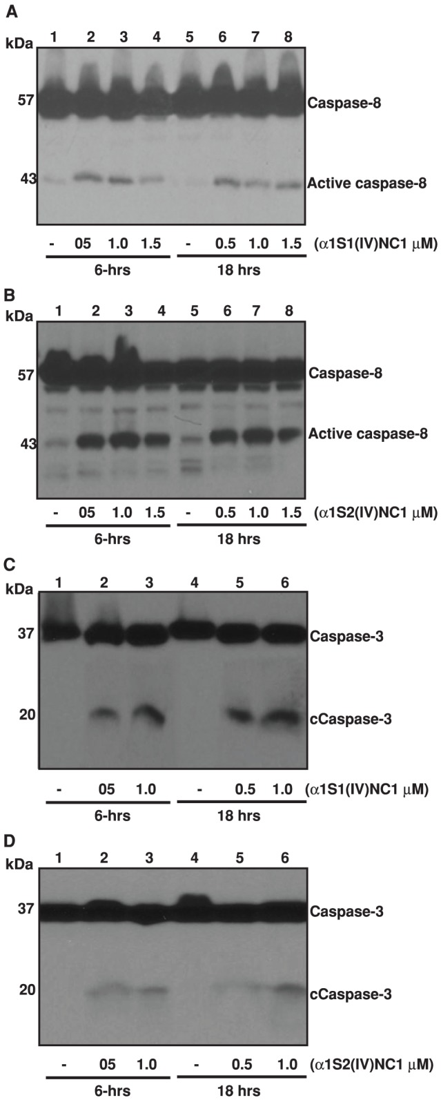
Figure 4. Caspase-8 activation (A and B).

Mouse choroidal endothelial cells (ECs) were incubated with and without different doses of α1S1(IV)NC1 and α1S2(IV)NC1 domains for 6 and 18-hrs, and total cells lysed for 30-min in ice-cold RIPA lysis buffer and about 25 µg of cytosolic extract per lane was separated and immunoblotted with primary antibodies against caspase-8. Caspase-3 activation (C and D). ECs were incubated with and without different doses of α1S1(IV)NC1 and α1S2(IV)NC1 domains and total cytosolic extract immunoblotted with primary antibodies against caspase-3.
