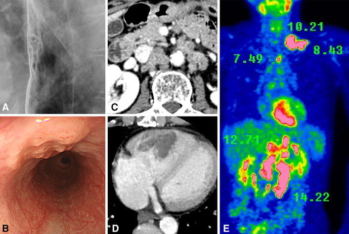
Fig. 1.

Esophageal squamous cell carcinoma was detected in the middle thoracic esophagus (a, b). Contrast-enhanced CT showed the multiple para-aortic lymph node metastases and cardiac right ventricle metastasis (c d). FDG-PET showed multiple high accumulations of FDG (shows SUV counts) (e)
