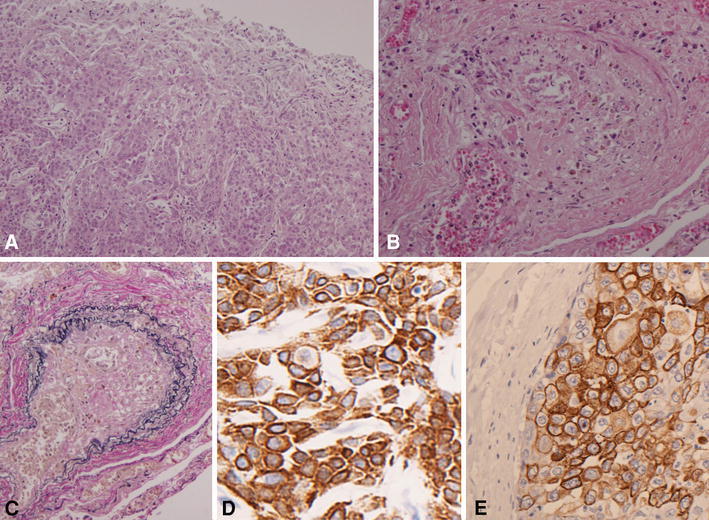
Fig. 3.

Microscopic examination. The primary tumor of the esophagus (a H&E ×100). Multiple tumor emboli and clot formation were shown in the pulmonary arterioles (b H&E ×100). EVG staining of the pulmonary arterioles showed concentric fibrocellular intimal proliferation with narrowing of the lumen (c EVG ×100). Immunostaining for CK5/6 proved that both the positivity rate and staining intensity were high (d primary tumor of the esophagus, CK5/6 ×400, e tumor cells in the pulmonary arteriole, CK5/6 ×400)
