
FIGURE 1.
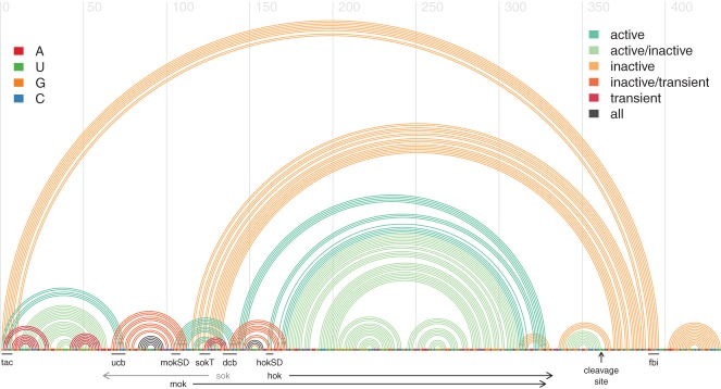
RNA structure features for the reference sequence from E. coli plasmid R1 encoding the hok and mok proteins. The horizontal line depicts the plasmid's sequence with its nucleotides color-coded according to the legend on the top left. Underneath the sequence line, black arrows indicate the protein-coding regions of the hok and mok proteins. The gray arrow shows the sequence region that is complementary to the sok anti-sense RNA, which is part of a different transcript. Each arc above the horizontal line represents a base pair between the two corresponding positions along the sequence and is color-coded according to the structure conformation to which it belongs (active, inactive, or transient; see the legend on the top right). Below the horizontal sequence line, black lines indicate the location of known sequence motifs: (tac) translational activator element; (ucb) upstream complementary box; (dcb) downstream complementary box; (mokSD) mok Shine-Dalgarno sequence; (hokSD) hok Shine-Dalgarno sequence; (fbi) fold-back inhibitory element. This arc-diagram was first published by Steif and Meyer (2012) and generated using the R-chie web server (Lai et al. 2012).