Figure 1.
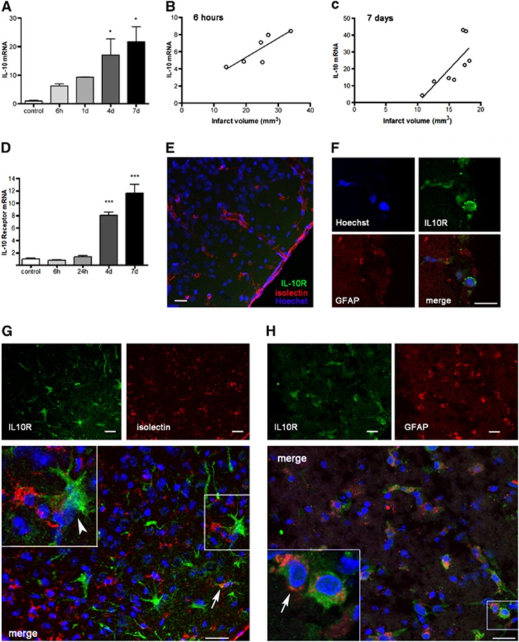
Ischemia upregulates the expression of interleukin-10 (IL-10) and IL-10 receptor (IL-10R) in wild-type (WT) mice. (A) The expression of IL-10 mRNA progressively increases in brain tissue after permanent middle cerebral artery occlusion (pMCAO) from 6 hours (h) to 7 days (d). (B, C) The extent of IL-10 mRNA expression correlates (linear regression analysis, P<0.05) with infarct volume that was measured by magnetic resonance imaging (MRI) (T2) in the same animals 6 hours (B) or 7 days (C) after pMCAO. (D) Increased expression of IL-10R is apparent from 4 days after ischemia, but not within the first day. mRNA values are expressed as fold increase versus control nonischemic brain tissue. (E to H) Immunofluorescence staining of IL-10R (green) in the cortex of controls (E) and after pMCAO (F to H). (E) IL-10R immunoreaction (green) is not detected in the control cortex. Isolectin (red) staining shows resident microglia and blood vessels. (F) Isolated IL-10R-positive cells are detected in the subarachnoid space on the top of the ischemic cortex after pMCAO. (G) IL-10R immunoreactive cells (green) are seen in superficial cortical layers from 4 days after ischemia. These cells are rarely positive (arrow) for isolectin (red) and most of the IL-10R+ cells have morphology (arrowhead) compatible with that of reactive astrocytes. (H) Double staining with GFAP (red) shows that most of the IL-10R+ cells are astrocytes (arrow). The cell nuclei were stained with Hoechst (blue). Images in (F to H) correspond to days 1, 4, and 7 after pMCAO, respectively. Bar scale: 20 μm. n=4 to 6 mice per group. *P<0.05 and ***P<0.001.

