Figure 2.
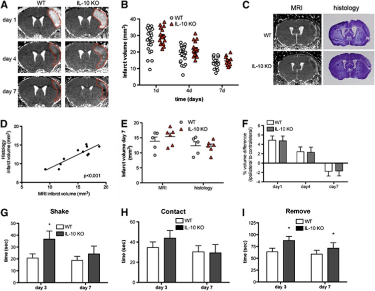
Interleukin-10 (IL-10) deficiency slightly increases infarct volume and neurologic deficits. (A) Brain infarction was longitudinally monitored by T2 magnetic resonance imaging (MRI) in the same mice at days 1, 4, and 7 after ischemia and a representative animal per group is shown. (B) Infarct volume is slightly larger in IL-10 knockout (KO) mice (n=17) than in the wild-type (WT) (n=20) (two-way ANOVA by genotype and time; P<0.05 for genotype effect; P<0.001 for time effect). (C) In an additional group of mice, MRI was followed by histology in the same mice. T2 maps of representative brain slices at day 7 after permanent middle cerebral artery occlusion (pMCAO) are shown with their corresponding histologic staining (cresyl violet). (D) Infarct volume measured by MRI and by histologic means in the same mice shows a good correlation (linear regression analysis, r2=0.8, P<0.001). (E) The infarct volume measured by histologic means is significantly smaller (P<0.001) than the corresponding MRI measures. However, with either method, genotype group differences at day 7 are not statistically significant. (F) At day 1, the ratio of the volume of the ipsilateral to the contralateral hemisphere is above 1 due to edema. The effect is attenuated at day 4, whereas at day 7 the ipsilateral hemispheric volume is reduced. However, hemispheric volume differences between WT and IL-10 KO mice are similar along time. (G to I) Measure of the time to shake (G), contact (H), and remove (I) steps of the adhesion/removal tape test shows worse neurologic deficits at day 3, as shown by a significant delay in the time to shake (G) and remove (I) for the contralateral (left) forepaw, but the worsening effects of IL-10 deficiency are attenuated at day 7. Animals in (F to I) are the same as in (B), *P<0.05.

