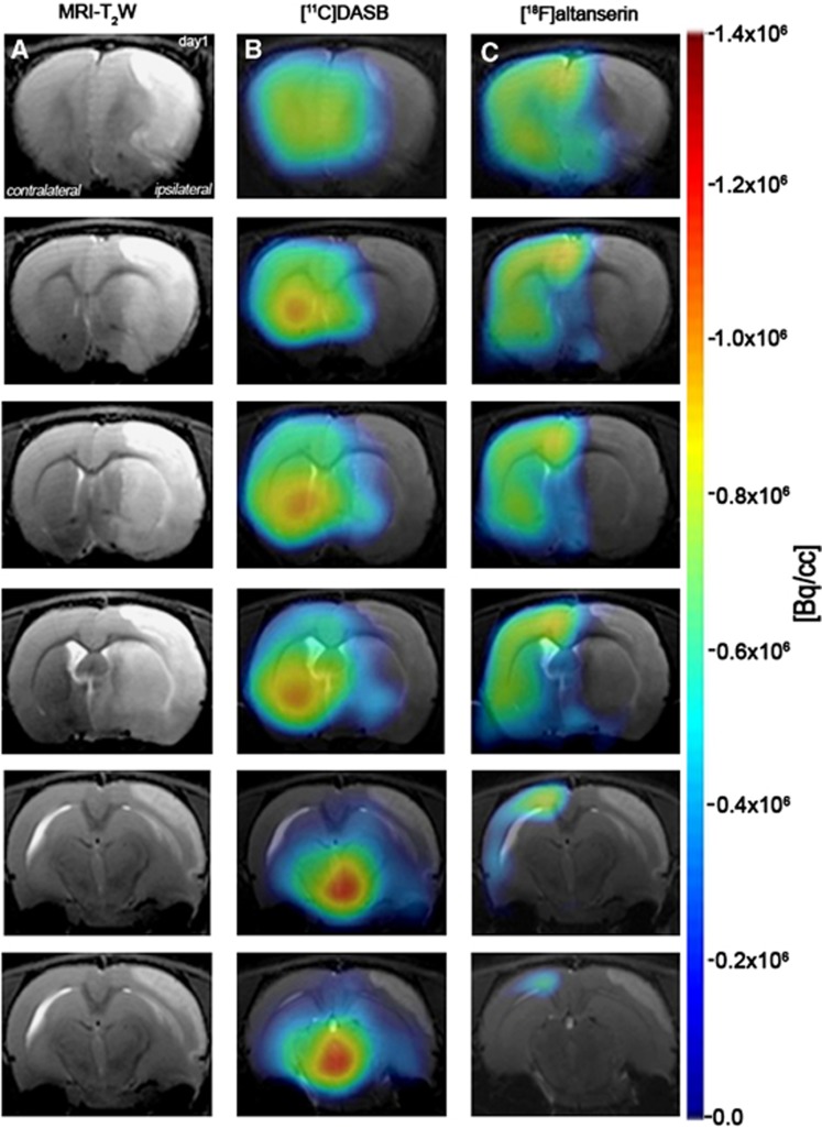
Figure 2.
Magnetic resonance imaging (MRI) (T2-weighting (T2W)) and positron emission tomography (PET) images of [11C]DASB and [18F]altanserin at day 1 after cerebral ischemia. Serial MRI (T2W) (A), serotonin transporter (SERT) (B) and 5-HT2A PET binding (C) images of axial planes at the level of the lesion. PET images are coregistered with the MRI (T2W) of the same animal to localize the PET signal. Images correspond to the anterior–posterior representative brain slices covering the extension of the lesion.

