
Figure 2. Expression of PD-L1, PD-L2 and PD-1 in ID8 mice.
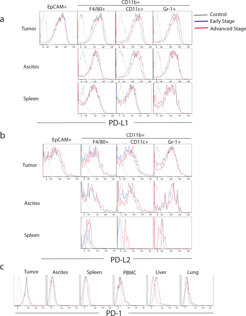
Mice were inoculated i.p. with 5×10⁶ ID8 tumor cells (n=12), and their tumor, ascites, spleen, liver, lung and blood were harvested from half of the mice at early and the other half at advanced tumor. Tumor cells (EpCAM+), macrophages (CD45+CD11B+F4/80+), DCs (CD45+CD11c+) and MDSCs (CD45+CD11B+Gr1+) were isolated from the tumor, ascites and spleen. Histograms show PD-L1 (a) and PD-L2 (b) expression by ID-8 tumor cells as well as macrophages, DCs and MDSCs from tumor, ascites and spleen of ID-8 tumor-bearing mice. (c) PD-1 expression on CD8+ T cells from tumor, ascites, spleen, blood, liver and lung of ID-8 tumor-bearing mice. Results are from one of the 3 experiments.