
Figure 4. PD-1 or PD-L1 blockade increases immune activation of TILs in ID8 tumor.
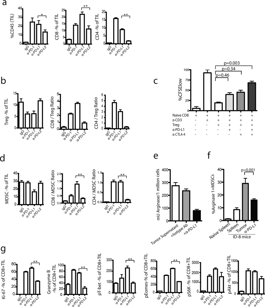
One week following completion of blockade treatment, the TILs were harvested from regressing tumors and stained with various markers. Percentages of CD8+ and CD4+ TIL (CD45+) infiltration of total leucocytes (a) and the ratio of CD8+ T cells to Tregs (b) are shown in treated versus untreated groups. (c) Blocking PD-1/PD-L1 interaction reduced Treg-mediated suppression of CD8+ T cells in vitro. CFSE-labeled CD8+ T cells were co-cultured with syngeneic, αCD3-loaded DCs with or without Tregs and αPD-L1 or αCTLA-4 as indicated. CD8+ T cells and stimulator APCs were obtained from naive B6 mice. Tregs were obtained from ID8-tumor bearing mice. Treg-mediated suppression of proliferation of naïve CD8+ T cells was noticed. Results from one of 3 experiments are shown. (d) The ratio of CD8+ T cells to MDSCs are shown. (e) CD8+ TILs from αPD-L1 treated mice were stained with arginase-1 (8C9 clone from Santa Cruz) and analyzed by flow cytometry. The CD11b+arginase-1+ MDSCs within CD45+ TILs are shown. (f) Tumor-dervived MDSCs were plated at 1×10⁶/well in 24-well plates and stimulated with equal amount of tumor supernatants (from ID8 cells). Following stimulation, cells were added with αPD-L1 and then arginase I was analyzed after 24 h following washing with PBS and lysis buffer. (g) Percentage of Ki67+ and Granzyme B+ as well as MFI of pT-bet, pEomes, pS6, and pAkt expression by CD8+ TILs are shown. The results are the sum of three independent experiments with 8–10 mice per group.