Figure 1.
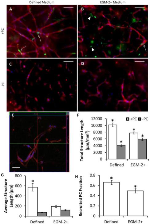
Vasculogenesis in 96-well plate constructs. (A-D) Representative images of whole mount CD31 stains of HUVECs (red) with (A-B) or without (C-D) PCs (green) in defined medium (A,C) or EGM-2+ (B,D) after 3 days of culture. Nuclei are stained blue. Arrows indicated recruited PCs, and arrowheads indicated PCs that were not recruited. Scalebars = 100 μm. (E) Confocal z-stack of a construct similar to that of (A). The large image is one slice of the stack, and the top and right images represent optical cross-sections through the sample at the green and red lines. Scalebar = 30 μm. (F-H) Quantitation of microvessel network properties. HUVECs grown with PCs in defined medium demonstrated improved total structure length (F), average structure length (G), and PC recruitment (H) over all other conditions. *p < 0.05 in comparison to all other conditions.

