Figure 6.
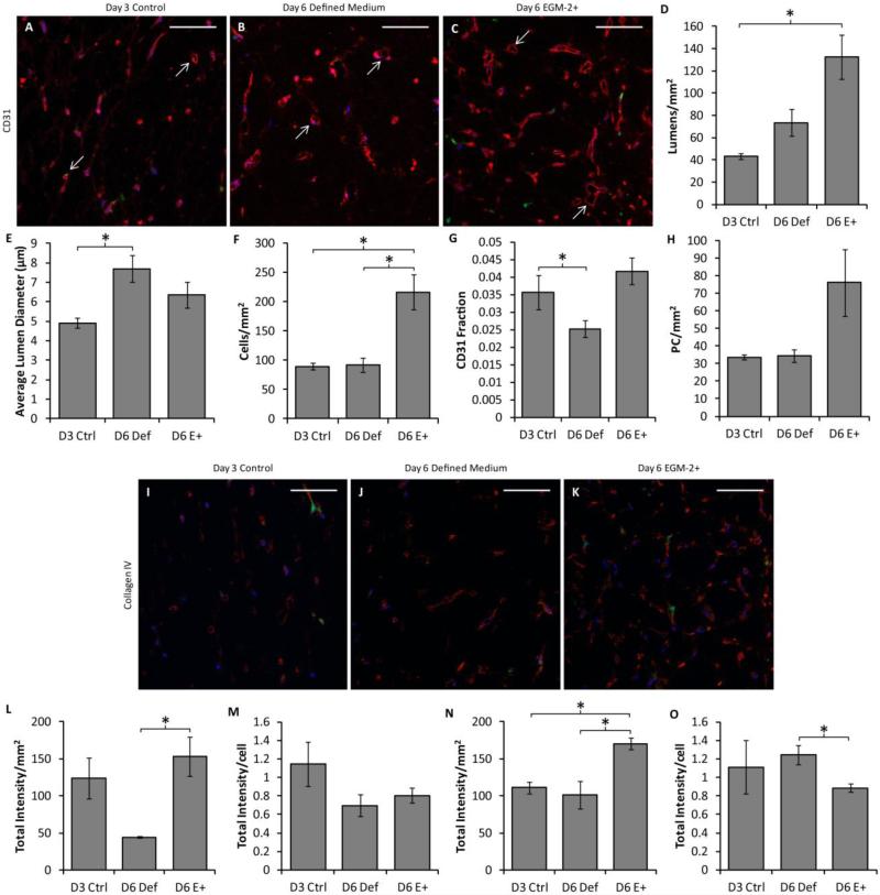
Analysis of the hybrid approach based on cell densities and basement membrane deposition. CD31, laminin and collagen IV staining of slab cross sections was performed. (A-C) Representative CD31 staining (red) of slabs cultured in defined medium for 3 days (A), 6 days (B) or defined medium for 3 days followed by EGM-2+ for 3 days (C). PCs are green and nuclei are blue. Arrows indicate examples of lumens that were characterized. Scalebars = 100 μm. (D-H) Quantification of sections. The lumen density was improved in hybrid slabs over the control (D), and the average lumen diameter was larger in day 6 defined medium slabs (E). The cell density (F) was highest in the hybrid slabs. The area fraction of CD31 staining (indicative of EC density; G) was increased in hybrid slabs over day 6 defined medium slabs, but the PC density (H) was constant across conditions. (I-K) Representative collagen IV staining of control (I), defined medium (J), and hybrid (K) slabs. Laminin staining was similar and therefore is not shown. (L-O) Quantification of basement membrane staining. More total laminin staining was present per area in hybrid slabs than day 6 defined medium slabs (L); however no difference was observed in staining intensity per cell (M). A similar trend was observed for collagen IV staining, except that the staining intensity per cell was decreased in hybrid slabs (N-O).

