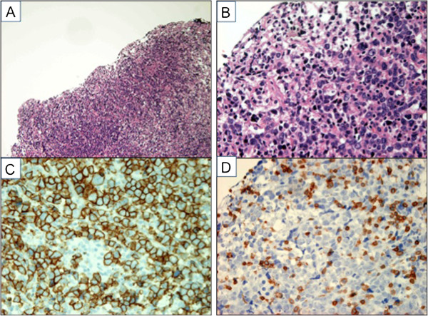
Figure 2.

Histology of the pituitary lesion. A. Hematoxylin-Eosin (HE) stain (×100). The pituitary gland is completely replaced by abnormal lymphocytes. B. HE stain (×400). The sections show diffuse proliferation of large-sized abnormal lymphoid cells. C. CD20 immunostaining (×400). These atypical cells are positive for CD20. D. CD3 immunostaining (×400). These atypical cells are negative for CD3.
