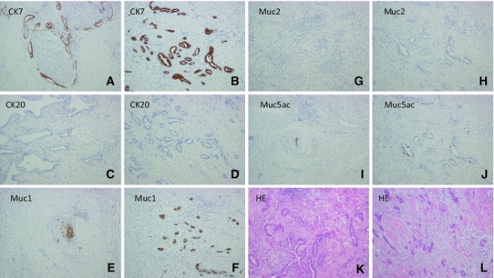
Fig. 2.

Immunohistochemical examination showed the staining pattern of the primary bile duct cancer to be the same as that of the thoracic tumor. Both the primary cancer (a, c, e, g, i, k) and thoracic tumor (b, d, f, h, j, l) were well stained by CK7 (a, b), CK20 (c, d), MUC1 (e, f), MUC2 (g, h), MUC5ac (i, j), and H&E (×100)
