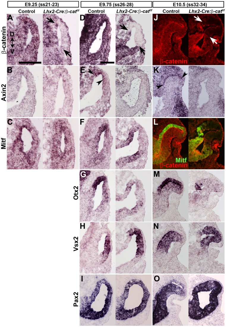
Figure 3. Canonical Wnt/β-catenin signalling is required for RPE cell commitment.
(A–C) In situ hybridization analyses of the indicated genes on coronal sections of E9.25 somite stage 21–23 (ss21–23) control embryos (left panels) and Lhx2-Cre:β-cateninflox/flox embryos (right panels). (D–I) In situ hybridization analyses of the indicated genes on coronal sections of E9.75 (ss26–28) control embryos (left panels) and Lhx2-Cre:β-cateninflox/flox embryos (right panels). (J–O) In situ hybridization analyses (K, M–O) or immunohistochemical analyses (J, L) for gene expression of the indicated genes on coronal sections of E10.5 (ss32–34) control embryos (left panels) and Lhx2-Cre:β-cateninflox/flox embryos (right panels). β-catenin protein is indicated by red labelling in J and L while Mitf protein is indicated by green labelling in L. Arrows indicate the area where β-catenin has been inactivated in A, D and J. Arrow heads indicate the area of Axin2 expression in E and K. Dorsal-ventral (D–V) orientation for all panels is indicated in A. Scale bars: (A–C and D–O) 100 µm.

