
Figure 1.
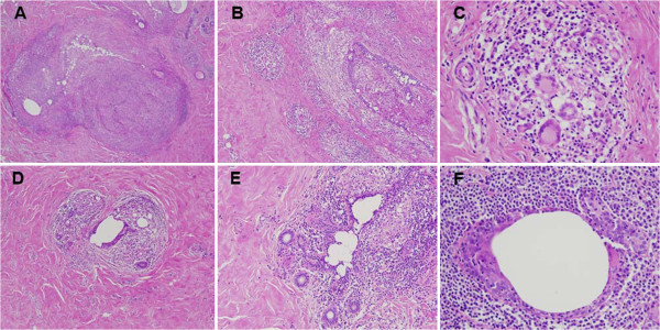
Histological sections of breast nodules examined after hematoxylin and eosin staining. (A) Chronic granulomatous inflammation of nodular arrangement (×40). (B and C) Multiple confluent granulomas rich in multinucleated giant cells (B ×100 and C ×400). (D and E) Lobulite lymphocytic and granulomatous (D ×100 and E ×200). (F) Periductal inflammation rich in neutrophils, macrophages and lymphocytes (×400).