
Figure 3.
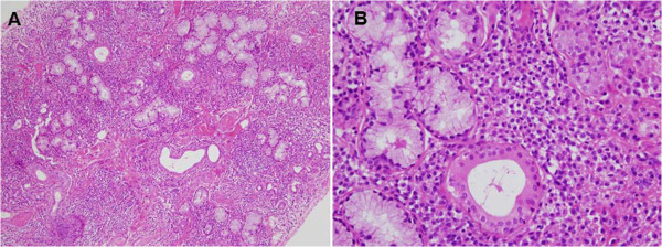
Histological biopsies of salivary glands examined after hematoxylin and eosin staining. (A) Note the presence of multiple inflammatory foci (focus: nodular clusters of at least 50 cells) associated with collagen fibrosis, corresponding to a level 4 according to the classification of Chisholm and Mason (×100). (B) It shows an intense infiltrate of chronic inflammatory lymphocytes and plasma cells, interstitial, especially surrounding the acini and the ducts (×400).