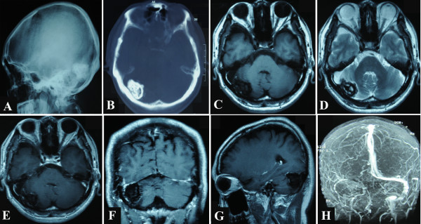
Figure 1.

Preoperative radiological images. Normal skull X-ray and unenhanced axial computed tomography (CT) scans display an irregular calcified intracranial lesion in the right occipital region, which is separated from the lamina interna cranii by a narrow cleft. Magnetic resonance (MR) images display a blur nodule in the right transverse sinus and the neighbouring dura. The lesion presents with mixed low and equal signal intensity on unenhanced T1-weighted image and mixed low and high signal intensity on unenhanced T2-weighted image. It exhibits irregular linear contrast enhancement only in its dural periphery after administration of gadolinium. Magnetic resonance venogram (MRV) reveals no detectable venous flow of the right transverse sinus and the right sigmoid sinus. A, Lateral normal skull X-ray films; B, unenhanced axial CT films; C, unenhanced axial T1-weighted MR image; D, unenhanced axial T2-weighted MR image; E, enhanced axial MR image; F, enhanced coronal MR image; G, enhanced sagittal MR image; H, MRV image.
