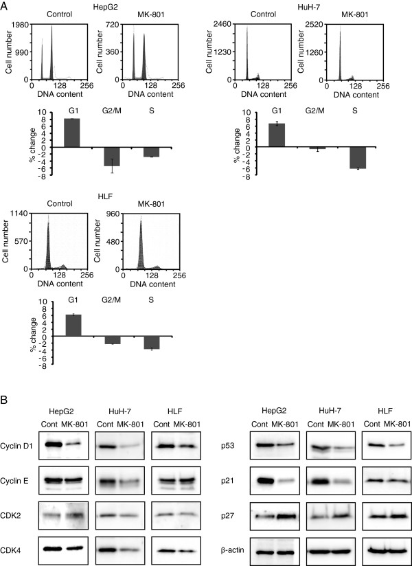
Figure 2.
Cell cycle analyses by flow cytometry and Western blotting. A: Hepatocellular carcinoma cells were cultured in 10-cm dishes with or without 250 μM MK-801 and were harvested after 72 h by trypsinization and fixed in 70% ethanol. Cells were incubated in 1ml PBS containing 50 μg propidium iodide and 200 μg RNase A. Flow cytometric analysis was performed with a FACSEpics XL flow cytometer. Each bar represents the mean ± SD of three replicates. B: Cell lysates from control or MK-801 (250 μM) treated cells were prepared and proteins were separated on SDS-PAGE gels and transferred onto nitrocellulose membranes. Western blot analysis was performed with indicated antibodies.

