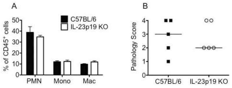
Fig. 6. IL-23 is not required for influx of acute inflammatory cells into the oviducts or the development of oviduct pathology.

(A) On day 10 post-infection, flow cytometric analysis revealed no difference in the frequency of innate inflammatory cells in the oviducts of C57BL/6 (black bars) and IL-23p19 KO mice (white bars). Bars represent the mean ± SEM of the frequency of CD45+ cells in the oviducts of 4 mice per strain for one representative experiment of two. PMN: Ly6G/Chigh F4/80negCD11cneg; Mono (inflammatory monocytes): Ly6G med F4/80negCD11cneg; Mac (macrophages): F4/80pos (B) Histological analysis of oviduct dilatation in genital tracts harvested on day 42 post-infection revealed no difference between the strains (P > 0.05 via Mann-Whitney U-test). Data points represent semi-quantitative scoring of oviduct dilatation of individual mice for 5 mice per strain from one representative experiment of two. C57BL/6 (black squares) and IL-23p19 KO mice (open circles). Median indicated by horizontal line.
