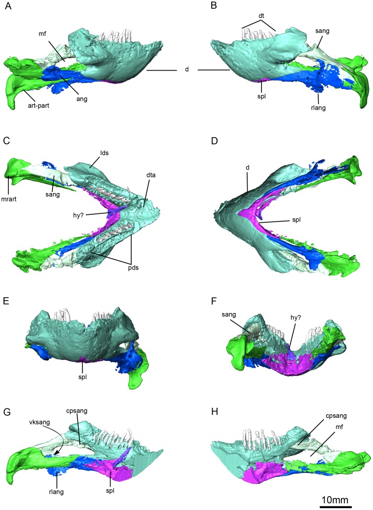
Figure 9. Niassodon mfumukasi mandible.
Left lateral (A), right lateral (B), anterior (C), posterior (D), ventral (E), dorsal (F) views; right ramus in medial view (G) and left ramus in medial view (H). ang, angular; art-part, articular-prearticular complex; cpsang, conical process of the surangular; dt, dentary teeth; dta, dentary table; hy?, probable hyoid; mrart, median ridge of the articular; lds, lateral dentary shelf; pds, postdentary sulcus; rlang, reflected lamina of the angular; sang, surangular; spl, splenial; vksang,ventral keel of the surangular.

