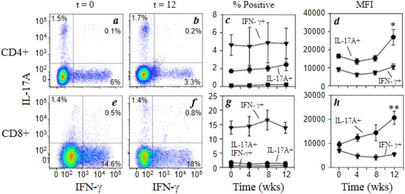
Fig. 2.
Chronic morphine exposure increases the functional activity of Th17 and Tc17 cells. Rhesus macaque PBMCs were stained for cell surface expression of CD3 (Alexa700), CD4 (Qdot605), and CD8 (Qdot655) followed by intracellular staining for IFNγ (FITC) and IL-17A (Alexa647). Representative data for Th1 and Th17 CD4+ cells (a, b), and Tc1 and Tc17 CD8+ cells (e, f) for animals at 0 week (a, e) and 12 weeks (b, f) of morphine administration are presented. The pooled results for the full macaque panel (7 animals) are presented as the percent positive IFNγ + (▼), IL-17A + (●) and IFNγ +IL-17A + (■) for CD4+ cells (c) and CD8+ cells (g). The level of intracellular cytokine expression was also determined for the CD4+ (d) and CD8+ subpopulations (h) and is expressed as mean fluorescence intensity (MFI). Data are presented as the mean (± sem) for the 7 animals. *p<0.05; **p<0.01

