Fig. 4.
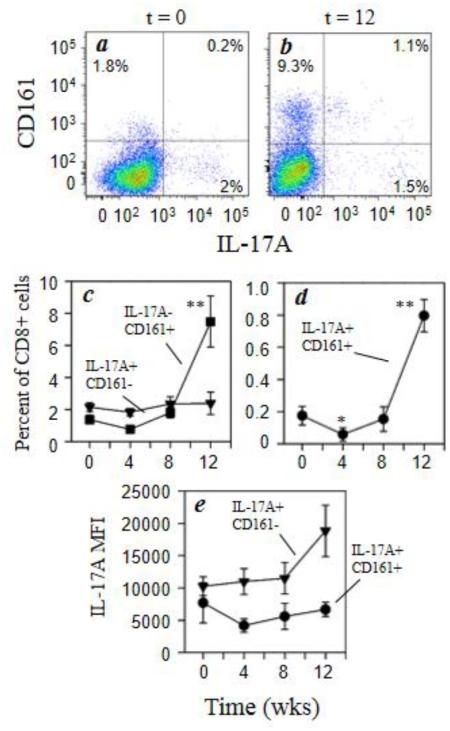
Morphine increases expression of CD161 by CD8+ T cells. Rhesus macaque PBMCs were stained for cell surface expression of CD3 (Alexa700), CD8 (Qdot655), and CD161 (PerCP-Cy5.5) followed by intracellular staining for IL-17A (Alexa647). Representative data for the co-expression of CD161 and IL-17A within the CD3+CD8+ T cells (a, b) for animals at 0 week (a) and 12 weeks (b) of morphine administration are presented. The pooled results for the full macaque panel (7 animals) are presented as the percent positive IL-17A−CD161+ (■) and IL-17A+CD161− (▼) and IL-17A+CD161+ (●) for CD8+ cells (c, d). The level of intracellular cytokine expression was also determined for the IL-17A+CD161− (●) and IL-17A+CD161+ (▼) and is expressed as mean fluorescence intensity (MFI) (e). Data are presented as the mean (± sem) for the 7 animals. *p<0.05; **p<0.01

