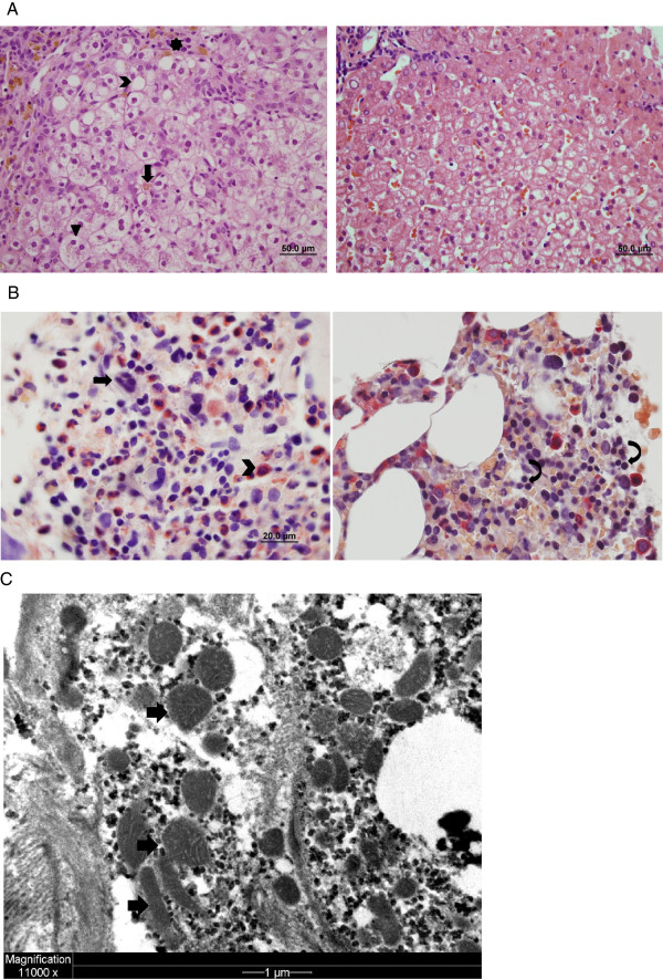
Figure 1.
Liver and bone marrow pathology. A: The patient’s bone marrow (left photo) contains megakaryocytes (arrow) and numerous myeloid cells (chevron), while erythroid cells are difficult to identify. In contrast, erythroid cells (curved arrows) are readily apparent in control bone marrow (right photo). B: The patient’s liver (left photo) shows lobular disarray with hepatocyte ballooning (arrowhead), canalicular cholestasis (arrow) and zone 1 steatosis (chevron). Iron deposition (star) is present in macrophages and hepatocytes. Control liver (right photo) for comparison. C: Electron micrograph of hepatocyte shows mild pleomorphism of mitochondria (arrows), which contain predominantly flattened, straight and curved cristae. These are non-specific findings that can be seen in normal liver.

