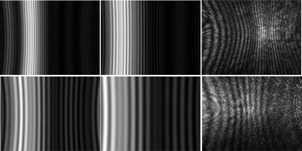
Fig. 5.
The projection images calculated from angle-resolved scattered light distribution by the Mie theory with Θ=24° for images in the left column and Θ=16° in the middle column. The images in the right column are measured diffraction images of the spheres with x=200µm. 1st row: d=25µm; 2nd row: d=9.6µm.

