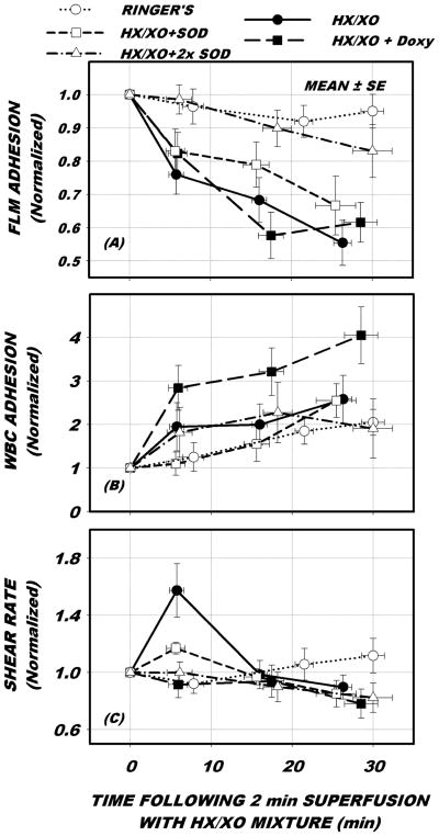
Figure 1.
Venular time course of adhesion of lectin coated fluorescently labeled microspheres (FLMs) and leukocytes (WBC), and wall shear rate in response to baseline conditions (○, Ringers alone), following 2 min exposure to mixture of hypoxanthine and xanthine oxidase (•, HX/XO), HX/XO exposure preceded by and following the addition of 0.5 μM doxycycline (■, HX/XO + Doxy), and HX/XO preceded and followed by superfusion with super oxide dismutase (SOD) at 1X (□) and 2X (Δ) concentrations. All data were normalized with respect to control period conditions given in Table 1. As shown in panel A, FLM adhesion fell significantly 45% in 30 min following exposure to HX/XO and was not affected by the addition of Doxy. Introduction of SOD significantly mitigated FLM shedding, in a dose dependent manner. WBC-EC adhesion (panel B) did not rise significantly in response to HX/XO. With the addition of SOD, WBC-EC adhesion rose significantly with SOD at 1X concentration, but not with SOD at 2X. Doxycycline caused a significant rise in WBC-EC adhesion. Shear rates (panel C) remained mostly constant with all treatments, except for a transient rise in response to HX/XO within the first 5 min.

