Figure 5.
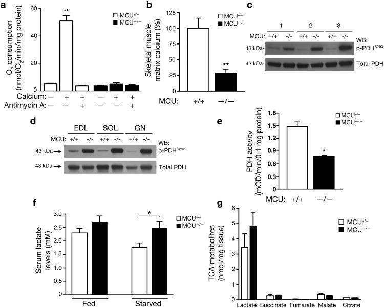
Altered in vivo skeletal muscle metabolism and PDH activity in MCU-/- mice. a) Mitochondrial oxygen consumption following depolarization induced by the addition of 500 μM calcium with and without the respiratory chain inhibitor Antimycin A (15 μM). The average +/- SEM of three independent experiments is shown. **p<0.01 by t-test compared to without calcium. b) Levels of matrix calcium measured in WT and MCU-/- mitochondria derived from skeletal muscle following an overnight 16 hour fast (**p<0.01 by t-test; n=3 WT and N=4 MCU-/- mice). c) Western blot determination of the levels of phospho-PDH (serine 293 of the E1-α subunit) and total PDH levels in the skeletal muscle of three pairs of WT and MCU-/- mice starved for 16 hours. d) Under starved conditions, altered PDH phosphorylation is seen various muscle types including the extensor digitorum longus (EDL) representing glycolytic/fast twitch fibers, the soleus (SOL) that is predominantly oxidative/slow twitch and the gastrocnemius (GN) that is a mix of fast and slow twitch. e) Skeletal muscle PDH activity in WT and MCU-/- mice after a 16 hour fast (*p< 0.05; n=3 mice per genotype). f) Serum lactate levels in WT and MCU-/- male mice under fed conditions or after 16 hours of starvation (n=7 WT and n=6 MCU-/-, *p<0.05 by t-test). g) Metabolomic analysis of skeletal muscle demonstrating levels of various TCA cycle intermediates in mice that were starved overnight (n=3 per genotype). There was a trend for increased lactate levels in the MCU-/- muscle. All pooled data represents mean +/- S.E.M.

