
Figure 1.
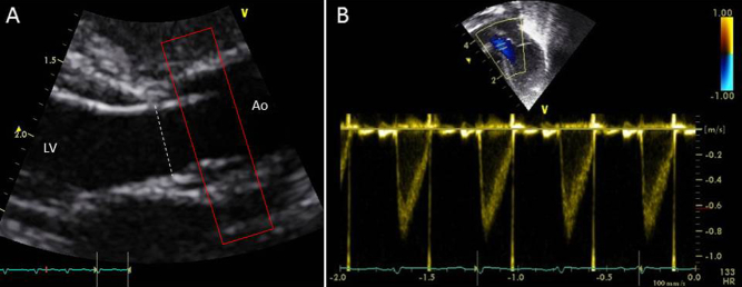
Echocardiographic quantification of LVO, with (A) diameter measured from the parasternal long-axis view and (B) velocity assessed from the apical view. Approximate location of PC MRI acquisition plane is also demonstrated (red box). Ao, Aorta; LV, left ventricle.