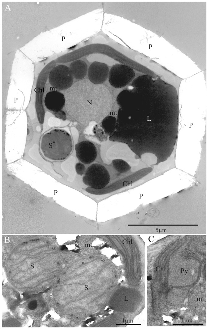
Figure 2. TEM images of Braarudosphaera bigelowii specimens -A and -B.
(A) B. bigelowii specimen-B from offshore Tomari port, Tottori, showing nucleus (N), chloroplasts (Chl), lipid globules (L), pentaliths (P), mitochondria (mt) and spheroid body (S). (B) B. bigelowii specimen -A from Tosa Bay, Kochi, Japan, showing detail of spheroid bodies (S). Note that the structure contains about 10 lamellae. The chloroplast (Chl) and lipid globules (L) can also be seen. (C) Detail of chloroplast of B. bigelowii specimen -A from Tosa Bay, Kochi, Japan, showing a bulging type of pyrenoid (Py). The mitochondrial profile (mt) can be seen.

