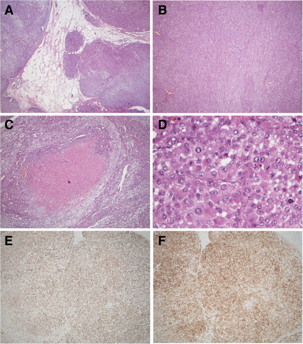
Figure 2.
The pathological findings of the tumor showed (A) sinusoidal and capsular invasion with irregular border (hematoxylin and eosin, 40×), (B) sheet-like arrangement (40×), (C) central necrosis (100×), (D) high grade nucleus with frequent mitoses (400×), and positive immunohistochemical staining for both (E) melan A and (F) alpha-inhibin.

