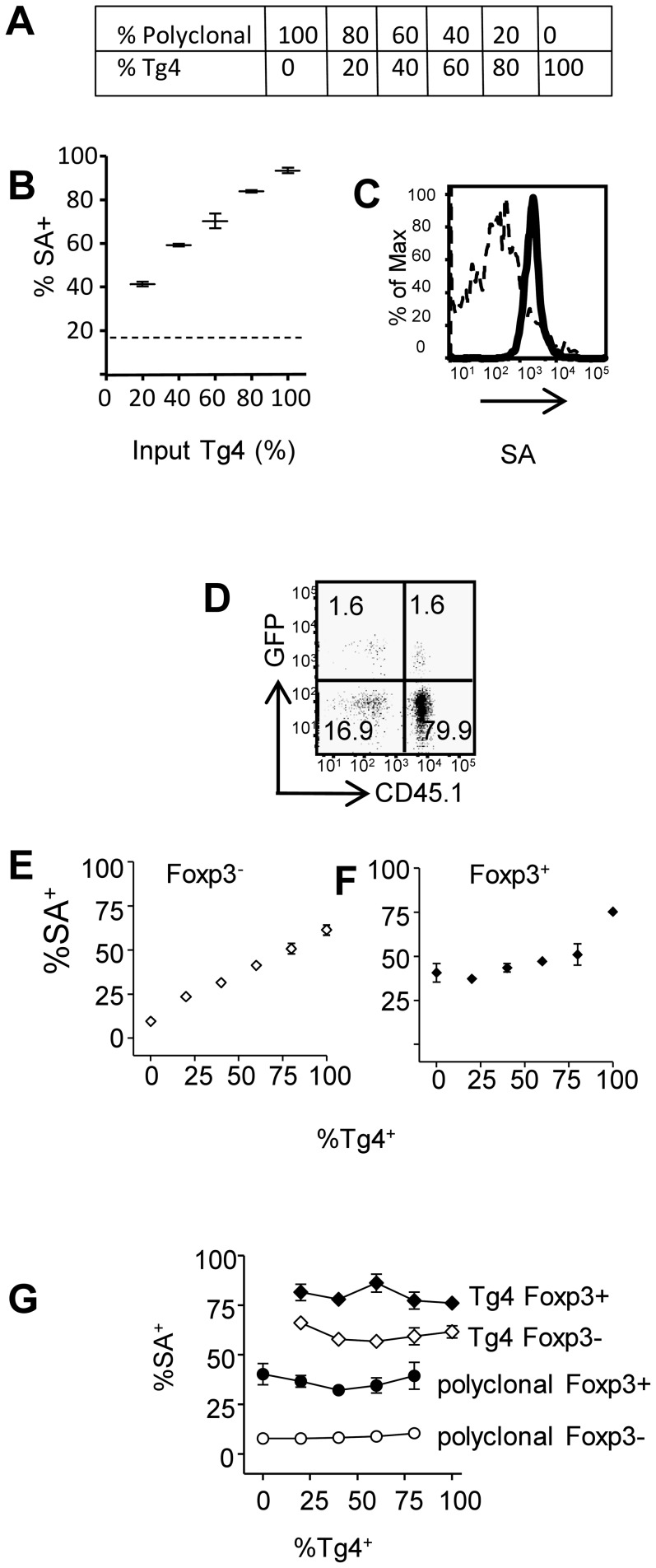
Figure 2. Detection of antigen reactive conventional and regulatory T cells by membrane transfer in mixed populations.
(A–C) MBP-reactive Tg4 T cells (CD45.1+) were mixed with polyclonal B10.Pl CD4+ T cells (CD45.1−) at the ratios indicated in (A) and were co-cultured with 4Tyr-pulsed biotinylated APC. (B) Detection of membrane transfer by staining with streptavidin-APC (gating on all CD4+ T cells) the dashed line indicates the background streptavidin-APC staining in the absence of any Tg4 T cells. (C) Streptavidin-APC staining of polyclonal (dashed line) and Tg4 T cells (solid line) after co-culture. (D–G) Tg4-Luci-DTR-4 T cells (CD45.1+) were mixed with polyclonal Foxp3-GFP CD4+ T cells (CD45.1−) and co-cultured with 4Tyr pulsed APC as in (A). (D) Gating of GFP− and GFP+ polyclonal and Tg4 CD4+ T cells (80% Tg4 : 20% polyclonal). Detection of membrane transfer within the Foxp3− (E) and Foxp3+ (F) CD4+ populations with increasing frequency of TG4 T cells. (G) Membrane transfer within Foxp3− (open symbols) and Foxp3+ (closed symbols) Tg4 (and polyclonal T cells across the range of Ag-reactive : bystander T cells tested. Error bars show SD. Data shown is from one experiment representative of 2 similar experiments.

