
Figure 2.
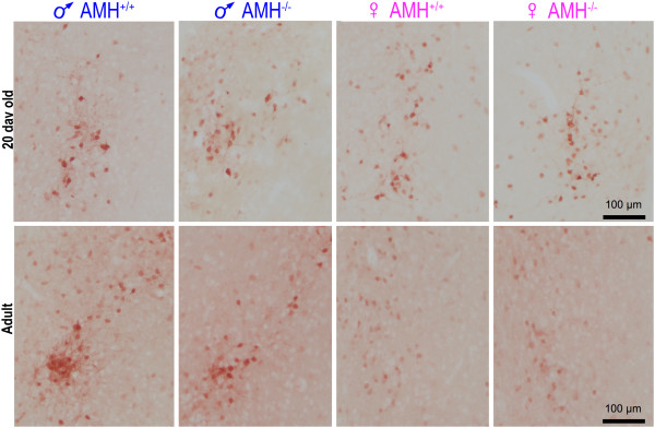
The dimorphism in the CALB-SDN varies with age and Amh genotype. The images are photomicrographs of the CALB-SDN illustrating the appearance of the nucleus in pre-pubescent (20 days old) and adult mice. All sections were stained with anti-calbindin antibodies. The location of the CALB-SDN relative to the anterior commissure and third ventricle is illustrated in Figure 1, with the quantitative estimates of the number and size of the calbindin+ve neurons illustrated in Figures 3 and 4, respectively. ♂, male; ♀, female.