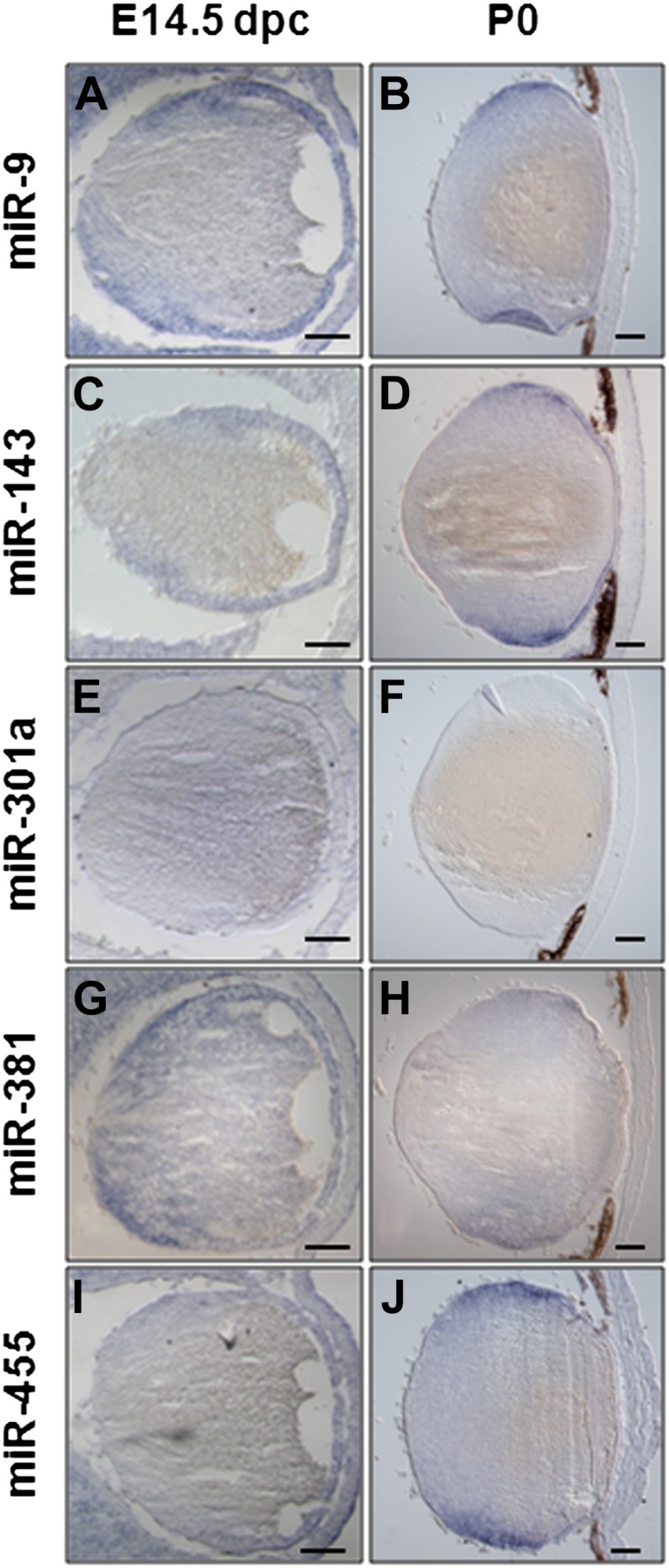
Figure 11.

Analysis of miR-9, -143, -301a, -381, and -455 expression pattern during embryonic and postnatal lens development. (A to K′) RNA in situ hybridization on frontal eye sections of wild-type mice at different developmental stages (as indicated in the panels) were hybridized with specific probes. At E14.5, the miR-9 (A), -143 (C), -301a (E), -381 (G), and -455 (I) expression domain included the monolayer of lens epithelial cells, the proliferating lens cells, the migrating lens cells, and the differentiating lens cells. Note for miR-301a (E), a weak staining is detectable in these structures at E14.5. At postnatal day P0, the distribution of both miR-9 (B) and miR-143 (D) is largely maintained in all the lens cells previously described for the E14.5 lens, whereas miR-301a (F) is not detected. At this stage both miR-381 (H) and miR-455 (J) expression domains are restricted to the equatorial zone of the lens in proliferating, migrating, and differentiating lens cells.
