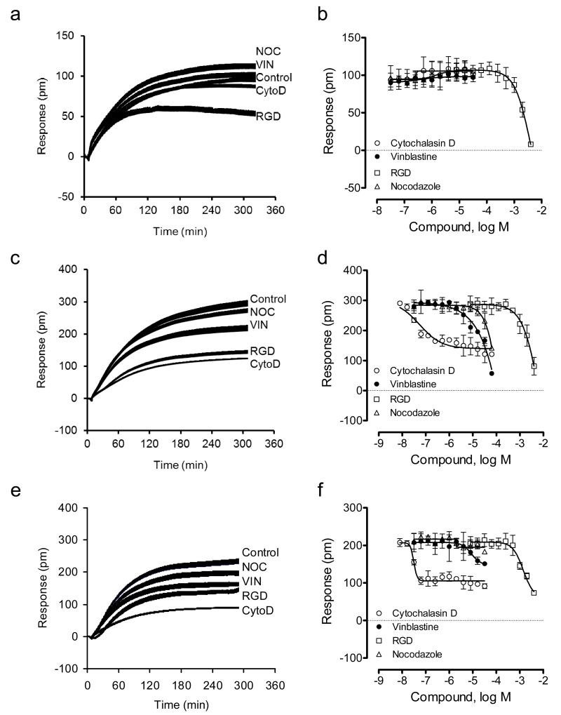
Figure 4.
DMR characterization of HEK-β2AR-GFP cell adhesion in the presence of different inhibitors under ambient condition. (a-f) The DMR of the engineered cells adherent onto different sensor surfaces: tissue culture treated (a, b), fibronectin coated (c,d), and collagen IV coated (e,f). (a,c,e) real-time DMR; (b,d,f) the DMR amplitudes at 5hr after cell seeding as a function of inhibitor dose. For all, the total number of cells added were the same; that is, 18,000 cells per well. For (a,c,e), the inhibitor dose was fixed to be 10μM, 10μM, 10μM and 1mM for nocodazole, vinblastine, cytochalasin D, and RGD peptide, respectively. The data represents mean±s.d. (n=12 for a, c, and e; n =4 for b, d, and f).

