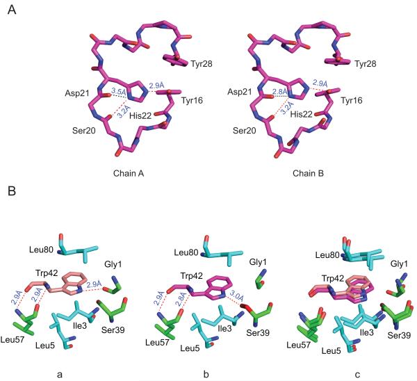
Figure 2. Local conformations in the P23T and HGD γD-crystallin structures.
(A) No difference in His22 side chain conformations is observed in the two independent protein molecules in the asymmetric unit. (B) Contacts around the Trp42 side chain in the HGD (PDB ID: 1HK0) (a) and the P23T (b) structures and their best-fit superposition (c).

