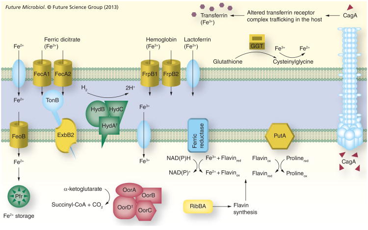
Figure 1. The iron sources available in the stomach and the ferric uptake regulator-mediated responses aimed at maintaining the intracellular iron balance in Helicobacter pylori.
Yellow color indicates that expression is inactivated by iron-bound ferric uptake regulator (Fur). Green color indicates that expression is inactivated by apo-Fur. Red color indicates that expression is activated by iron-bound Fur. The subcellular location of GGT, PutA, HydABC and ferric reductase is only for illustration and has not been experimentally determined.
†Iron–sulfur proteins.
CagA: Cytotoxin-associated protein; GGT: γ-glutamyltranspeptidase; Pfr: Bacterial ferritin.

