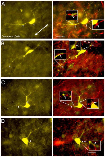
Figure 5.
Fluorescence photomicrographs showing contacts between FluoroRuby-labeled cortical boutons and FluoroGold-labeled disc-shaped or stellate commissural cells in the central nucleus of the left IC. The first column shows commissural cells in the left IC retrogradely labeled with FluoroGold (FG, yellow). The second column shows an overlay of the FG label and cortical axons labeled by FluoroRuby (FR, red) in the same field of view. Arrows indicate contacts between FG-labeled commissural cells and FR-labeled cortical boutons. Inserts show expanded views (3x enlargement) of contacts between commissural cells and cortical boutons. The orientation of the fibrodendritic laminae – from dorsomedial to ventrolateral (A, line with double arrows) - could be identified by the orientation of many of the labeled structures (axons and cells). Disc-shaped cells (A, B) had a somatodendritic orientation parallel to the laminae. Stellate cells (C, D) had no apparent orientation. All images were taken at the same orientation: dorsal is up and medial is to the right. Transverse sections. Scale bar = 20 μm.

