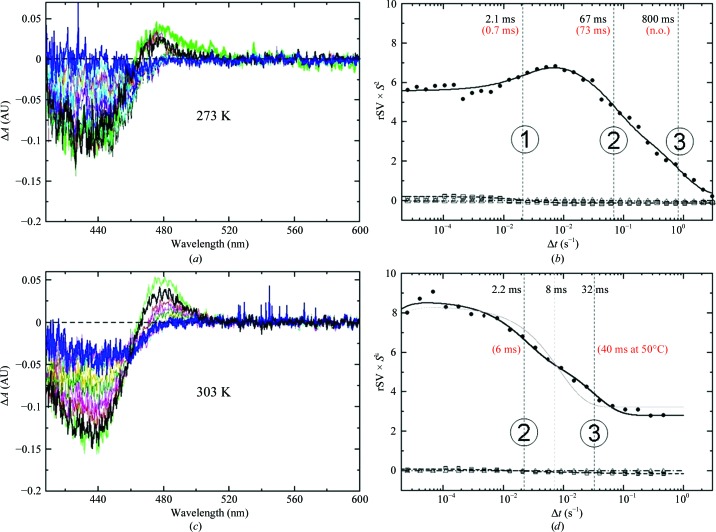
Figure 6.
Time-resolved difference absorption spectra of crystalline slurry at 273 K (a) and 303 K (c) (black, earliest time point; blue, last time point). (b, d) Right singular vectors (rSVs) from SVD analysis of the difference spectra. Solid lines, global fit by sums of three exponentials identifying three processes. Processes 1–3 are labeled with their respective relaxation times and are marked with thin vertical lines; relaxation times from crystallography are shown in red brackets (n.o., not observed). The gray line in (d) is the fit of the final relaxation with only one exponential.

