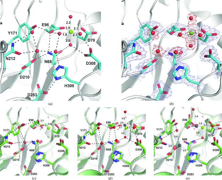
Figure 1.
New crystal structure of human APE1 with the native Mg2+ cofactor and previous structures with surrogate metals. (a) Close-up view of the active site of the new structure, showing Mg2+ and several ordered water molecules (molecule A of the three molecules in the asymmetric unit for PDB entry 4lnd). Also shown are the important catalytic residues; all but Asp70 are strictly conserved in the DNase I superfamily (Tyr171 is replaced by His in some members). The octahedral coordination of Mg2+ is indicated by dotted lines with distances provided (also given in Table 3 ▶). Hydrogen bonds are indicated by dashed lines. (b) The same view of the active site showing a 2F o − F c OMIT map contoured at 1.5σ for protein and waters and an F o − F c OMIT map contoured at 8.0σ for the Mg2+ ion (black mesh). Note that one or more of the three non-Mg2+-coordinating water molecules are not observed in the other two protein molecules (B and C) in the asymmetric unit. (c) Previously reported structure of Sm3+-bound APE1 (green; PDB entry 1bix; Gorman et al., 1997 ▶) aligned with the new Mg2+-bound structure (white). The coordination of the Sm3+ ion (cyan) is shown (dotted lines) with distances. The coordination of Mg2+ (green) in the new structure is also indicated (without distances). Water molecules shown as red spheres and hydrogen bonds (dashed lines) are for the Sm3+-bound structure. The water molecules that coordinate Mg2+ are shown as red stars. (d) The previously reported structure of APE1 with one Pb2+ ion (green; PDB entry 1hd7; Beernink et al., 2001 ▶) aligned with the new Mg2+-bound structure (white). The coordination of the Pb2+ ion (gray) is shown (dotted lines) with distances and the coordination of Mg2+ (green) is also indicated. Water molecules (red spheres) and hydrogen bonds (dashed lines) are for the Pb2+-bound structure (waters that coordinate Mg2+ are shown as red stars). (e) The previously reported structure of APE1 with two Pb2+ ions (green; PDB entry 1e9n; Beernink et al., 2001 ▶) aligned with the new Mg2+-bound structure (white). The coordination of the Pb2+ ions (gray) is shown (dotted lines) with distances and the coordination of Mg2+ (green) is similarly indicated. Water molecules (red spheres) and hydrogen bonds (dashed lines) are for the Pb2+-bound structure (waters that coordinate Mg2+ are shown as red stars).

