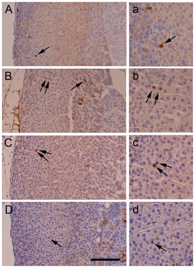
Figure 1. BrdU pulse-labelled cells in adult mouse adrenal glands.
(A-D) BrdU immunostaining of adrenal sections from female F1 mice (A) 4 hours; (B) 1 week; (C) 3 weeks & (D) 5 weeks after a single BrdU injection. Typical BrdU-labelled cells (brown nuclei) are indicated by arrows. (a-d) Higher magnification views of BrdU-positive nuclei that are shown in A-D. Scale = 100 µm in A-D; 50 µm in a-d.

