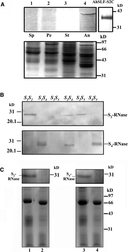
Figure 3.
Coimmunoprecipitation of AhSLF-S2 and S-RNases.
(A) Detection of AhSLF-S2 in several tissues and E. coli expressing AhSLF-S2C by a polyclonal antibody against the C-terminal region of AhSLF-S2. Bottom panel: Coomassie blue–stained gel before protein gel blot analysis showing control loading of proteins. Lanes 1 to 4 represent protein extracts from sepal (Sp), petal (Pe), style (St), and anther (An) from an S2S5 line. AhSLF-S2C indicates the detection of AhSLF-S2C in E. coli expressing AhSLF-S2C.
(B) Immunoblot analysis of style proteins from several lines of an S-allele–segregating family using allele-specific antibodies against peptides derived from S2- and S4-RNases. Genotypes are indicated at the top.
(C) Extracts of pollinated styles of S1S4 (lane 1) or S2S5 (lane 3) lines (50 μg) and pollen of an S2S5 line (50 μg) were immunoprecipitated with anti-AhSLF-S2-C antibody (3 μL) (lanes 1 and 3) or control preimmune serum (3 μL) (lanes 2 and 4), respectively. After immunoprecipitation, the proteins were subjected to 12% SDS-PAGE followed by protein gel blotting with specific antibodies against S2- and S4-RNases, respectively (top panels). S-RNases detected are indicated. Bottom panels: Coomassie blue–stained gels before protein gel blot analysis showing equal loading of proteins. The strongest bands represent immunoglobulin heavy chains. Molecular mass markers are indicated in kilodaltons.

