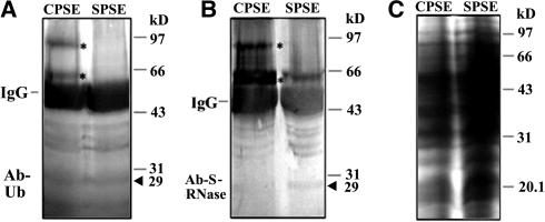
Figure 9.
Detection of Ubiquitinated S-RNases.
The pollen total proteins (50 μg) after incubation with CPSE or SPSE were immunoprecipitated with anti-S-RNase antibody (3 μL). The precipitated proteins were subjected to 12% SDS-PAGE followed by protein gel blot analysis with the anti-ubiquitin antibody (A) and the anti-S-RNase antibody (B). The 25 μg of total proteins also were subjected to 12% SDS-PAGE and stained by Coomassie blue for loading control (C). Immunoglobulin heavy chains are indicated by IgG. Asterisks indicate two extra protein bands detected by both the anti-ubiquitin antibody and the anti-S-RNase antibody after the incubation with CPSE. By contrast, no similar proteins were detected after the incubation with SPSE. Arrowheads indicate the predicted size of S-RNases.

