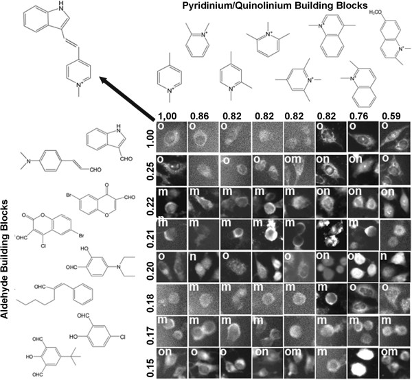
Figure 7.
Visualizing the relationship between the staining patterns of a reference probe (located at the upper left corner of the array; probe structure indicated by arrow) and the staining patterns of compounds with different chemical structures. Different aldehyde building blocks were plotted in rows (left) and pyridinium or quinolinium building blocks were plotted in columns (top). Numbers correspond to the Tanimoto coefficients between the different building blocks and the building blocks of the reference compound at the upper-left most corner of the array. As in Figure 6, individual cells representing the staining patterns observed in each image were manually cut from the images, and labeled based on their apparent organelle (o), membrane (m) or nuclear (n) staining patterns. Cells from images lacking significant signal or ambiguous in localization patterns were not labeled.

