
Figure 3.
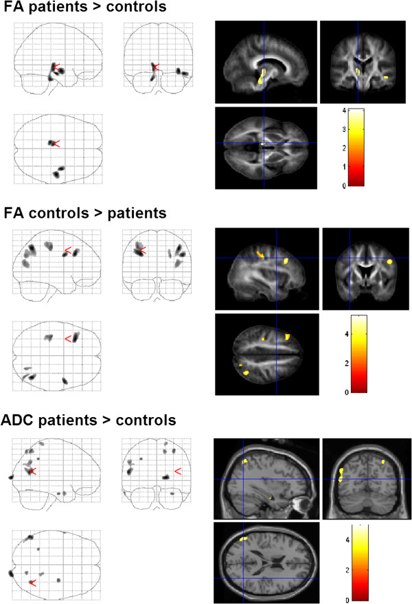
Changes of DTI-values in patients with dystonia compared to those in healthy controls (group comparison). FA was increased in brainstem, thalamus, and temporal areas. Decreased FA was observed in postcentral gyrus, frontal and occipital cortex. Diffusivity was enhanced in the area MT, and secondary somatosensory cortex.