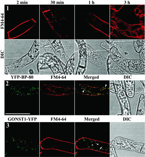
Figure 14.
Colocalization of YFP-BP-80–Labeled PVCs with Internalized FM4-64–Marked Organelles in BY-2 Cells.
Panel 1, uptake process of FM4-64 in living cells. Shown are steps of FM4-64 uptake profiled in living BY-2 cells in which the dye is first detected on the plasma membrane, followed by localization in internalized endosome-like structures and eventually insertion in the tonoplast. Panels 2 and 3, colocalization of PVC reporters with internalized FM4-64–labeled organelles. At 30 min after uptake, FM4-64–marked organelles colocalized with YFP-labeled PVCs in cells expressing the YFP-BP-80 reporter (panel 2) but remained separate from YFP-labeled Golgi stacks in cells expressing the GONST1-YFP reporter (panel 3). Bar = 50 μm.

