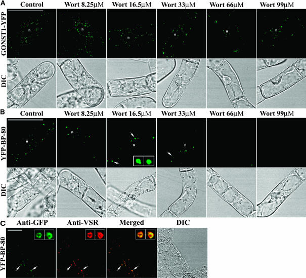
Figure 8.
Wortmannin Induces the YFP-BP-80–Labeled PVCs to Form Small Vacuoles in a Dose-Dependent Manner.
(A) and (B) Transgenic cells expressing the Golgi (A) and PVC (B) reporters were incubated with wortmannin (Wort) at various concentrations as indicated for 3 h before the treated cells were sampled for YFP imaging. Arrows in (B) indicate small vacuoles derived directly from the YFP-BP-80–labeled PVCs. The insets in the third panel of (B) are enlarged vacuole images of the two structures indicated by the arrows.
(C) Colocalization of anti-VSR (red) with PVC-derived vacuoles in transgenic cells expressing the YFP-BP-80 reporter after treatment with wortmannin at 16.5 μM for 3 h. The insets are enlarged vacuole images of the two structures indicated by the arrows.
n, nucleus. Bar in (A) and (B) = 50 μm; bar in (C) = 20 μm.

