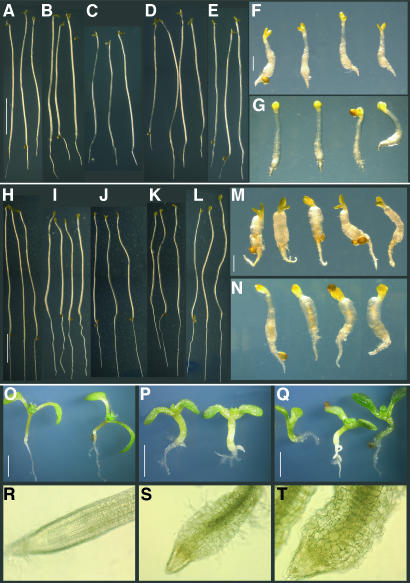
Figure 5.
Seedling Phenotypes of rcn1 pp2aa2-1 and rcn1 pp2aa3-1 Mutants.
Seedlings were grown for 7 d in the dark on standard medium containing no sucrose ([A] to [G]) or 1% sucrose ([H] to [N]), or for 7 d ([O] to [Q]) or 5 weeks ([R] to [T]) in the light on medium containing 1% sucrose. Wild-type Ws ([A] and [H]), Columbia ([E], [L], and [O]), and single mutant rcn1 ([B] and [I]), pp2aa2-1 ([C] and [J]), and pp2aa3-1 ([D] and [K]) seedlings show elongated hypocotyls, whereas rcn1 pp2aa2-1 ([F], [M], and [P]) and rcn1 pp2aa3-1 ([G], [N], and [Q]) show radial expansion and reduced hypocotyl and root elongation. Light microscopy reveals regular cell layers in rcn1 root tips (R) and dramatic cell expansion in rcn1 pp2aa2-1 (S) and rcn1 pp2aa3-1 (T) root tips. Scale bars = 5 mm in (A) to (E) and (H) to (L); 1 mm in (F), (G), (M), and (N); and 2 mm in (O) to (Q).

