Figure 1.

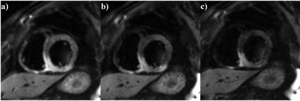
Short axis view of the inferior wall edema. CMR was performed 2 days after myocardial infarction. Diffusion weighted imaging with a) b = 50 mm/s2, b) b = 100 mm/s2, c) b = 200 mm/s2 acquired in a short axis plane.

Short axis view of the inferior wall edema. CMR was performed 2 days after myocardial infarction. Diffusion weighted imaging with a) b = 50 mm/s2, b) b = 100 mm/s2, c) b = 200 mm/s2 acquired in a short axis plane.